In this Medical Monday series, we’re talking about how your doctor reaches a diagnosis of common conditions associated with coagulation. It may help if you first read the introduction, before reading about today’s topic of heart valve conditions. Altogether we’re covering

- Introduction
- DVT, Deep Vein Thrombosis
- Post-thrombotic syndrome
- PE, Pulmonary Embolism
- Pulmonary hypertension
- AF, Atrial Fibrillation
- Heart Valve conditions – aortic and mitral
- Thrombophilia
- Stroke
Introduction
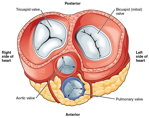 This picture of a cross-section across the heart shows your four heart valves.
Diseases of the heart valves are reasonably common particularly in older people. Disease most commonly affects the aortic or mitral valves. The aortic valve is the main valve out of the heart. The commonest problem is narrowing of the valve preventing the normal flow of blood from the heart. About half a million people in the US have severe aortic stenosis and more than 67,000 valve replacements are carried out each year. The mitral valve is the valve between the upper and lower chamber of the left side of the heart. It can either leak or become too narrow. Narrowing is called mitral stenosis, this can happen later in life after having rheumatic fever when young. In New Zealand there is an unusually high incidence of rheumatic fever.
In this post I will look at aortic stenosis.
Your story
 The symptoms of aortic stenosis are quite variable and can gradually creep up on you. Some of them only come on with exercise, these can include central chest pain, episodes of dizziness and fainting. You might also notice your heart racing (palpitations). Many of the symptoms resolve on resting. Over time you can become breathless on exercise and develop swollen ankles. However remember that some people have virtually no symptoms but just get slower. Sometimes fainting episodes are the only symptom.
Questions your doctor may ask
Your doctor will ask about your activity and and what brings on the symptoms. If you are getting central chest pain your doctor will be concerned and will want you to have more investigations. Your doctor may already know that you have a problem with your heart valve. Many cases are due to a congenital problem with your heart valve; as a child this causes no problems but as you get older the valve becomes stiffer and causes symptoms. Your doctor will also want to know if you have a family history of heart disease or if you had rheumatic fever as a child.
What your doctor may find
 There are a few signs that medical students learn about aortic stenosis but in reality these are quite difficult to pick-up. These are a low volume pulse and a drop in blood pressure. But it can be difficult to estimate pulse volume as it is often low in old people, also the blood pressure can be normal or even high in people with aortic stenosis. However people with aortic stenosis usually have a heart murmur that your doctor will hear. It has some characteristic features.
Your doctor will also examine your ankles for swelling and examine your abdomen to feel for your liver, but these signs come on late with this condition.
Tests you may have
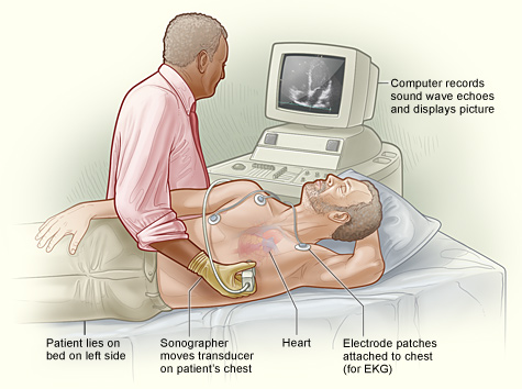 Echocardiogram: The most helpful investigation is an echocardiogram. With this your doctor can look at the movement of the aortic valve and measure the severity of the condition. An echocardiogram is also used o monitor your treatment.
ECG: The ECG measures the electrical activity of the heart and can help to assess the size of the left ventricle, which can become thickened as it is trying to pump blood through a very narrowed valve.
Chest X-ray: This can provide information about the size of the heart which can become enlarged with aortic stenosis. It might also show calcium deposits on the valve.
An exercise test: Some people with aortic stenosis have symptoms and get chest pain on exercise, but many have no symptoms. Your doctor may arrange and exercise test to see how well your heart manages under exercise. The ECG tracing during the exercise can provide information about performance.
Cardiac catheterisation: This is a more invasive test to look at the action of your heart. In this test a small catheter (a fine tube) is threaded through an artery up to the heart and a dye is injected while X-ray video is taken. The catheter is often insert through an artery in the groin. You might have this done before surgery as it shows if any of the arteries around your heart are blocked or narrowed, this problem can coexist with aortic stenosis.
What it could be
 Chest pain is a serious problem and your doctor will always want to investigate this further if there is any possibility of heart disease.
Dizziness and fainting can be due to abnormal heart rhythms, but is often far less serious and due to ear problems or sometimes just simple faints.
Palpitations are a common symptom. Many people have these intermittently, they are due to the occasional extra heart beat. They can be induced by anxiety or drinking coffee, but persistent palpitations or palpitations with other symptoms such as feeling faint may be a sign of heart disease.

Leave a Reply