Why would I have an ECG?
An ECG is a fairly routine test so it may just be used to check you have no major heart issues. On the other hand it may be used to look for atrial fibrillation (AF) or a heart attack (blocked blood supply to some of your heart muscle). If you attend the hospital with chest pain of any sort, including a pulmonary embolism, you are likely to have an ECG.
What is an ECG?
 An ECG or electro-cardiogram is a reading of your heart’s electrical signals and although it sounds very similar, is not to be confused with an echo cardiogram (an ultrasound of your beating heart).
Your heart muscle uses electrical impulses to cause rhythmical contractions of the muscle to pump blood, otherwise known as your heart beat. An ECG measures the electricity between various electrodes placed on your chest and after going through a computer draws a trace of the activity on special graph paper.
It just measures your body’s own electrical impulses, it doesn’t treat your heart in any way.
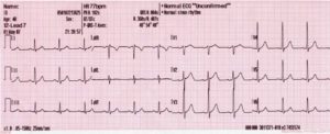
 the ECG leads – An ECG shows 12 slightly different heart tracings which are recorded between different pairs of electrodes;
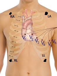
There are 4 limb electrodes, which are placed near the armpits and on the lower chest rather than actually on the limbs; RA, right arm; LA, left arm; RL, right leg and LL, left leg. The tracings from the limb electrodes are labeled I, II, and III and aVR, aVL and aVF
There are 6 pre-cordial (means in front of the heart) chest electrodes. The tracings from these are labelled V1 to V6.
These 12 tracings often only show 2 to 3 heart beats and it is the shape of the electric complex that doctors are interested in. Often at the bottom of the page will be a longer tracing (usually of trace II), which is used for checking the heart rhythm
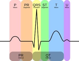
 the ECG waves – the electrical complex is labelled alphabetically from P to T. The P wave is the depolarisation of the atria; the QRS complex is the depolarisation of the ventricles and the T wave is the repolarisation of the ventricles.
The picture shows the wave form in trace II (the U wave is not usually seen).
What’s it like having an ECG?
An ECG is a very fast painless test; the most painful part can be pulling off the electrodes afterwards!
I just love the videos produced by Melbourne Children’s’ hospital and narrated by children. I don’t use these to be condescending in any way, I just find children’s perspective of tests to be very reassuring and down-to-earth. I especially like the bit in this video where it says “these pegs aren’t like the ones we use for the washing”.
What can an ECG show?
Heart rate and rhythm – The distance between each spikey QRS complex can be used to work out the heart rate and variations in distance between each complex is used to work out the rhythm. In atrial fibrillation, the heart rate is likely to be fast and the rhythm to be irregularly irregular. This is a medical term I love for its alliteration, but its used to distinguish the rhythm from one that is regularly irregular (for example; dropping a beat after each 3 heart beats in certain types of heart block)
Unusual depolarisation or repolarisation – the presence or absence of each wave and its shape help diagnose certain disorders. In AF there are no P waves because instead of having regular atrial depolarisation and contraction (which causes P waves) the atria are fluttering.
Damage to the heart muscle – the T wave and the ST segment (part between the S and T wave) are especially important in diagnosing angina or heart attack. Typically there is ST elevation, especially in the chest leads, soon after a heart attack.
Did you get to see your own ECG and have it explained to you?

Leave a Reply