Why would I have a heart echo?
Echos are used to examine your heart as it beats in real time, rather than just getting a one-time static picture. In relation to coagulation this could be needed to look for
- possible pulmonary hypertension (related to pulmonary embolism)
- underlying heart disease in atrial fibrillation,
- problems with your heart valves or
- to see how your heart is holding up after a massive pulmonary embolism.
It is an investigation to look at your heart and doesn’t alter or treat it in any way.
What is an echo and how is it related to dolphins?
 In last week’s post we said that a CTPA is basically X-rays taken to the extreme. Well, an echo is basically ultrasound taken to the extreme! Instead of X-rays, an echo uses sound-waves.
Dolphins can’t see clearly through murky water so instead of using their eyes to navigate they use their ears. They emit high-pitched clicks which bounce back to them when the sound hits a submerged object. They interpret this echo to work out where objects are. In the same way, medical ultrasound uses a probe to send out a much higher pitched sound and a computer to put together a visual image from the way the sound is bounced back. This is the same technology that is used for antenatal scans.
What’s it like having an echo?
I’ve found another great video in the series from the Royal Melbourne Childrens’ hospital. There’s something about a video narrated by a child that makes the whole process seem easy and down-to-earth! And this little boy has a wonderful Aussie accent!
Our son Isaac had plenty of heart echos as a baby before his open heart surgery. He didn’t even need sedation, so as an adult you should have no problems!
What can an echo show?
An echo can show three different aspects of your heart
1. heart structure
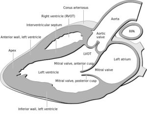
 The echo can show what the valves and heart walls look like. The technician will measure the thickness of the heart walls. This picture shows a cross-section of a normal heart in someone lying flat, as you would for an echo. You can see from this that the right ventricle is much smaller than the left ventricle with thinner walls – this is because the right ventricle is mainly a collection chamber for deoxygenated blood on the way to the lungs, whereas the left ventricle is a strong pump sending oxygenated blood all round the body.
In pulmonary hypertension the right ventricle has to push harder to overcome the high pressure in the vessels of the lungs. This means it becomes enlarged and the muscle walls become thicker (hypertrophied).
2. Any unusual blood flow in the heart
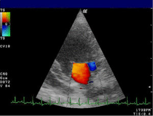
 Using doppler to show blood flow is a very pretty part of the examination, as it’s shown in colour. The technician can work out the direction and velocity of blood jets as the heart beats. This is very important in looking for congenital defects in babies. Usually, the left and right ventricles are totally separated with a muscular wall: In our son, Isaac’s case, the echo showed five small colourful jets of blood shooting through five holes in his heart (or in medical jargon – ventricular septal defects).
If doctors suspect a problem with your valves, they will look closely at the doppler pictures to see if there is any leakage (incompetence) or narrowing (stenosis). The picture shows blood flowing through the mitral valve.
3. Pressures and pumping strength of the heart
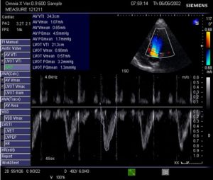
 An important measurement is the pressure in the pulmonary vessels. This should be very low (14 mm Hg) but gets much higher (above 25 mm Hg) in pulmonary hypertension.
Another important measurement is seeing what percentage of the blood in the left ventricle is pushed out with each heart beat. This is called the ejection fraction and is usually between 55% and 70%. If it below 40% the heart is too floppy to pump properly and if it is above 75% the heart is too stiff to pump properly. These both lead to heart failure. Regarding coagulation related conditions this is only likely to occur in heart disease in atrial fibrillation.
What health professionals are involved in an echocardiogram?
- A cardiac sonographer – usually this will be someone who has done radiography training (X-Ray) then undergone further training in ultrasound, then become a specialist in cardiac ultrasound
- a radiologist – a doctor specialised in all forms of imaging who will report the echocardiogram
- a cardiologist or cardio-thoracic surgeon – doctors specialised in heart medicine or surgery will also be part of the team and may do echos themselves.
What will an echocardiogram show in pulmonary hypertension?
Pulmonary hypertension leads to high pressure in the vessels of the lungs, right ventricular enlargement and wall thickening. In this rather specialised video there is a part at 1 minute 15 seconds showing this.

Leave a Reply