 Why would I have a CTPA?
If doctors are highly suspicious that you have a pulmonary embolism you will probably have a CTPA. It is an investigation to look for a PE and doesn’t alter or treat a pulmonary embolus in any way.
What does CTPA stand for?
Computerised tomography pulmonary angiography – quite a mouthful! We’ll consider the CT and PA components separately.
What’s CT?
- CT stands for computerised tomography. It’s basically X-rays taken to the extreme! Tomos is Greek for ‘slice’ and a doughnut shaped X-Ray machine takes multiple X-rays of different slices around your chest. It uses a series of narrow X-ray beams rather than the single beam used for a standard x-ray.
- The multiple single dimensional pictures obtained are then amalgamated by computer to give a 2D picture, which is far more detailed than a standard X-ray.
- CT is good for looking at soft tissues, blood vessels, and the brain.
What’s it like having a CT of the chest?
- You lie on a motorized couch, which goes into the scanner. There’s plenty of room (unlike an MRI scanner) so it’s unusual to feel claustrophobic. You keep still while the machine moves up and down the couch.
- This fantastic video, from Melbourne Children’s’ hospital, narrated by the cutest little girl, shows what fun this can be!
- Unfortunately, as an adult, you probably won’t be able to put a sticker on the scanner, and will have to wear a hospital gown, but otherwise the procedure is the same.
What’s PA?
- Pulmonary angiography means a picture of the vessels of the lung, which is where a pulmonary embolism lodges.
- You need an injection of a contrast dye to show the vessels more clearly.
What health professionals are involved in a CTPA?
- A radiographer/radiology technician will get you positioned correctly and perform your CTPA.
- A radiologist (doctor specializing in imaging) will give you the contrast injection and may well be present at your CTPA. The radiologist will interpret the CTPA and write a report. You can ask for a copy of this if you like.
Could you explain more about the pulmonary vessels?
 It is important to remember that a pulmonary embolus is not directly about the lungs, but about the vessels to the lungs. A clot forms in the deep veins of the legs (DVT) and breaks away, travels through the venous system to the right side of the heart, then out of the right ventricle to the pulmonary arteries where it becomes lodged.
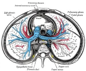
- The pulmonary arteries are shown in blue in this cross section of the chest, through the top part of the heart.
- It’s a bit confusing that the pulmonary arteries are coloured blue: We tend to think of arteries carrying red oxygenated blood and veins carrying blue deoxygenated blood.
- Yet vessels are actually named by their relationship to the heart; vessels that carry blood from the heart are called arteries and vessels that carry blood to the heart are called veins. Most arteries carry red oxygenated blood as they come from the left side of the heart after the blood has picked up oxygen from the lungs.
- However, the pulmonary arteries carry deoxygenated blue blood as they come from the right side of the heart and the blood has not yet reached the oxygen in the lungs. This means that although a PE occurs in the pulmonary arteries, it is one of the VENOUS thrombo-embolic disorders (VTEs) and very different from arterial clots like those in a heart attack.
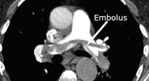
What does a pulmonary embolism look like on CTPA?
- Understanding a CTPA is a bit like looking at a prenatal scans and seeing a baby rather than a hazy picture of black dots! If you can do that, then you’ll probably be able to see that a CTPA looks very like the drawing of the pulmonary vessels above, although in back and white not colour!
- A radiologist explains a CTPA with pulmonary emboli in this video. You can see how the clot has a worm-like shape as it retains the shape of the leg vein where it was formed.
Can a CTPA pick up anything else as well as pulmonary emboli?
A CTPA may show that part of the lung has collapsed; atelectasis, or that there is a fluid collection in the lung lining,; a pleural effusion. Both these conditions can be associated with a PE, but they can also occur with chest infections.

Leave a Reply